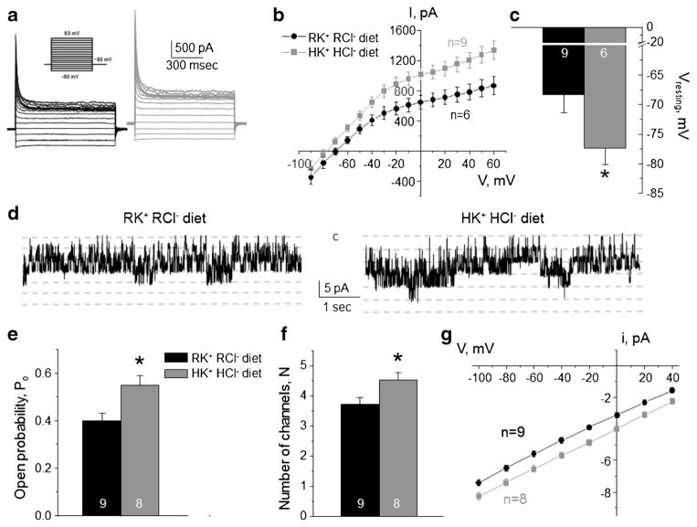
Fig. 2. High K+ high Cl− diet increases Kir4.1/5.1 activity on the basolateral membrane of collecting duct principal cells.
Representative macroscopic currents in response to voltage steps from − 90 to + 60 mV (shown in inset) from the holding potential of − 60 mV (a), averaged current-voltage (I–V) relations (b), and the summary graph of resting basolateral membrane potential (c) in principal cells from mice kept on regular diet (0.9% K+, 0.5% Cl−: RK+ RCl−, black) and high KCl diet (6% K+, 5% Cl−: HK+ HCl−, gray). d Representative continuous current traces from cell-attached patches monitoring basolateral 40 pS Kir4.1/5.1 single channel activity in collecting duct principal cells for the regular (left) and high KCl (right) conditions. Patches were clamped to −Vp = − 40 mV. “C” denotes non-conducting closed state. Summary graph of changes in open probability (e), number of active channels within a patch (f), and unitary current amplitude (g) of Kir4.1/5.1 in principal cells from mice on regular diet and high KCl diet. *Significant difference versus regular diet

