
Fig. 1.
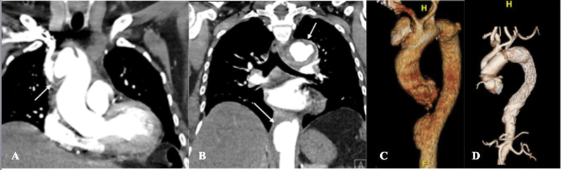
( A ) Admission computed tomography angiogram (CTA; coronal view) of patient 1 demonstrating 4.5 cm saccular pseudoaneurysm of distal ascending aorta and proximal aortic arch (arrow). ( B ) CTA (coronal view) of patient 1 with descending thoracic aortic pseudoaneurysms (arrows). ( C ) Preoperative three-dimensional reconstruction of patient 1 demonstrates multiple contained aortic ruptures and bovine aortic arch. ( D ) Postoperative three-dimensional reconstruction of patient 1 with intact ascending and descending aortic aneurysm repairs.