
Fig. 3.
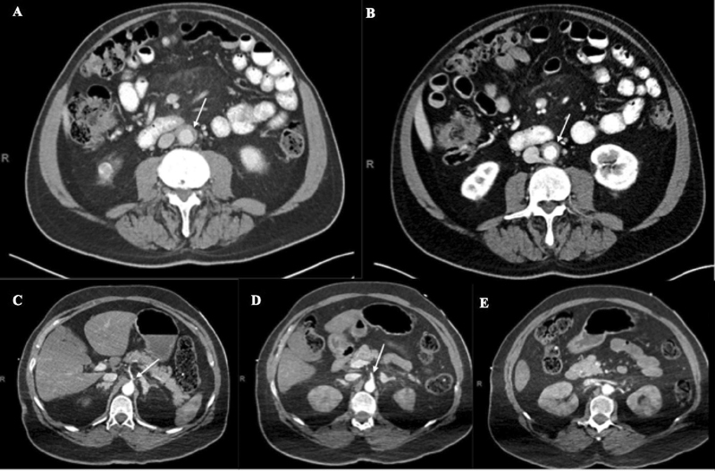
( A ) Computed tomography angiogram (CTA; axial view) of patient 3 demonstrating mild rind of soft tissue thickening surrounding infrarenal abdominal aorta (arrow). ( B ) Interval CTA (axial view) 3 months after ( A ) with unchanged rind of soft tissue thickening about the infrarenal abdominal aorta without aneurysm (arrow). ( C and D ) Admission CTA (axial view) of patient 4 demonstrating diminutive caliber of celiac trunk and superior mesenteric artery, respectively (arrow). ( E ) CTA (axial view) of patient 4 demonstrating hypoenhancement in bilateral kidneys.