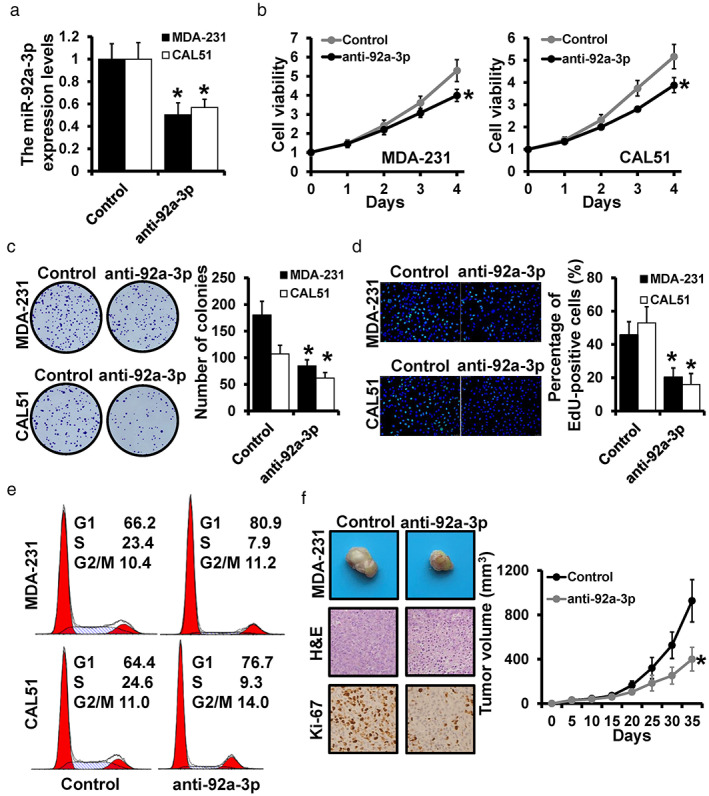
FIGURE 2.

miR‐92a‐3p‐depletion inhibits breast cancer progression. (a) The expression levels of miR‐92a‐3p in miR‐92a‐3p‐depleted CAL‐51 cells and MDA‐231 cells were detected by qRT‐PCR. For cell proliferation assay, MTT (b), colony formation assay (c), and EdU assay (d) were performed in miR‐92a‐3p‐depleted CAL‐51 cells and MDA‐231 cells. (e) The distribution of the cell cycle was detected by flow cytometry. (f) Representative photos and volumes of tumors, as well as H&E staining or Ki‐67 expression of xenograft tumors. *p < 0.05
