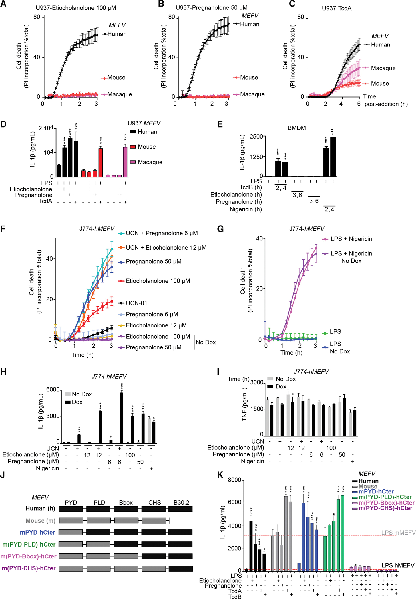
Figure 5. The human specificity of the response to steroid catabolites is intrinsic to the pyrin protein.

(A–C) Doxycycline-induced U937 monocytes or (D and K) PMA-differentiated U937 macrophages expressing human (black), mouse (red), Macaca fascicularis (magenta) or the indicated chimeric (J and K) MEFV were treated with the indicated stimuli.
(A–C) Propidium iodide (PI) incorporation was monitored every 5 min for 3–6 h.
(D and K) IL-1β concentration in the supernatant was quantified at 3 h (etiocholanolone, pregnanolone) or 6 h (TcdA, TcdB) post-addition.
(E) WT bone marrow-derived macrophages (BMDM) were primed for 16 h with LPS (100 ng/mL) and treated with TcdB (10 ng/mL), etiocholanolone (100 μM), pregnanolone (50 μM), or nigericin (10 μg/mL). IL-1β concentration in the supernatant was quantified at the indicated time point post-compound addition.
(F–I) J774 macrophages expressing or not (Dox or No Dox) human MEFV were treatedwith the indicated stimuli.
(F and G) PI incorporation was monitored every 5 min for 3 h.
(H and I) Cells were primed for 3 h with LPS before stimuli addition. (H) IL-1β and (I) TNF concentrations in the supernatant were quantified at 3 h post-addition.
(K) The dotted lines indicate the basal value in LPS-treated U937 cells expressing human MEFV or murine MEFV.
(A–I and K) One experiment representative of three independent experiments with mean and SEM of biological triplicates is shown. (D and E, H, I, and K) One-way ANOVA with Sidak’s test was used, ***p < 0.001 (H), *p = 0.214 (I), *p = 0.0304 (K), from left to right: *p = 0.0123, p = 0.0159.
