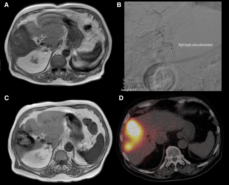
Fig. 2.
85 year old male diagnosed with hepatocellular carcinoma (HCC). At presentation, contrast enhanced T1 MRI (A), a solitary hypervascular lesion in segment 5, 6 and 8 with a maximum diameter of 8.1 cm was seen. At tumor board, the patient was considered for first-line SIRT. The 166Ho scout procedure consisted of a single injection of 233 MBq of 166Ho microspheres in the right hepatic artery (B) and subsequent SPECT/CT imaging showed no lung shunt, no extrahepatic deposition of activity elsewhere and visually good tumor targeting. The patient proceeded with 166Ho treatment in the afternoon (on the same day), in which 4.3 GBq of 166Ho microspheres were administered in the right hepatic artery (B). 3 months after treatment, follow-up contrast enhanced T1 MRI (C), showed a good response reducing its size from 8.1 cm to 5.8 cm and complete response according to mRECIST. Post-treatment SPECT/CT (D) 3 days after treatment confirmed the planned high accumulation of particles in the lesion, without extrahepatic deposition of activity (and no lung shunt). At this moment, more than 3 years after treatment, the patient has no signs of recurrent disease on imaging

