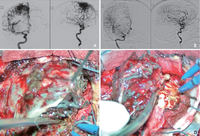
Fig. 1.
(A) Digital subtraction angiogram (DSA) before embolization, anterioposterior (AP) and lateral views. (B) DSA after embolizations and surgical excision, AP and lateral views. (C) Embolized vessels by Squid appeared black due to the admixed tantalum powders. (D) Circumferential dissection during surgical excision.

