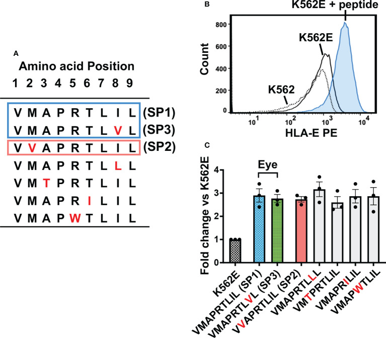
Figure 2.
HLA-E expression on K562E cells was comparable between UL40-derived signal peptides. (A) UL40-encoding signal peptides tested in this study are shown. The top two peptides (SP1, SP3) (boxed in blue line) were the predominant signal peptide sequences in the intraocular fluid, and the third peptide from the top (SP2) was detected in peripheral blood only (boxed in orange line). Amino acid substitutions compared to SP1 are indicated in red. (B) Representative histogram of HLA-E expression on K562, K562E cells cultured in the absence (K562E) or presence of peptides (VMAPRTLIL) (K562E+ peptide). (C) HLA-E expression on K562E cells pulsed with each peptide is shown. Data are shown as fold-change normalized to the mean fluorescence intensity (MFI) of HLA-E on K562E cells without peptide pulse. Representative data from three independent experiments are shown as means ± SEM. One-way ANOVA test was used for statistical analysis.

