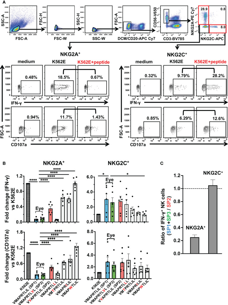
Figure 3.
UL40-encoding signal peptides identified in intraocular fluid strongly suppressed NKG2A+ NK cells. (A) Gating strategy for identification of two NK cell subsets, as determined by NKG2A+NKG2C- (NKG2A+) and NKG2A-NKG2C+ (NKG2C+), and representative flow cytometry plots of staining for IFN-γ and CD107 in NKG2A+ or NKG2C+ NK cells cultured in medium alone (medium) or with K562E cells (K562E) or K562E cells pulsed with peptides (VMAPRTLIL) (K562+peptide). (B) Inhibitory and activating effects of the seven representative peptides on NKG2A+ (two panels on left) and NKG2C+ (two panels on right) NK cells were compared. Data are shown as fold-change normalized to the response against unpulsed K562E cells. One-way ANOVA followed by Dunnett’s multiple comparison test. (C) Magnitudes of NKG2A-mediated inhibition and NKG2C-mediated activation by SP1 and SP3 were compared with those by SP2. Shown are ratios of anti-SP1 and -SP3 responses of NKG2A+ and NKG2C+ NK cells to anti-SP2 responses. The means ± SEM are shown. Representative data obtained for healthy CMV-seropositive individuals (n = 6) are shown. *P < 0.05, ****P < 0.0001.

