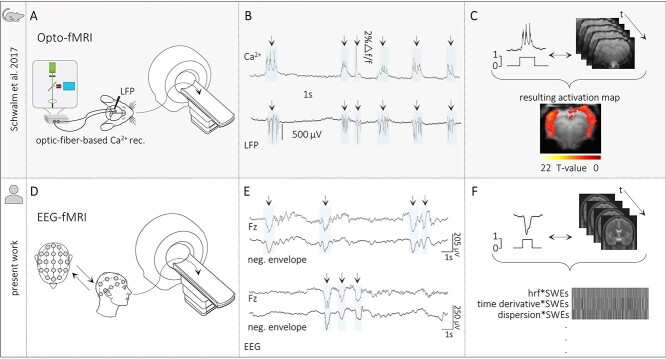
Figure 1.
Translational approach for SWE-related fMRI analyses in humans. (A) Scheme of opto-fMRI setup enabling simultaneous fiber-based calcium and fMRI recordings in anesthetized rodents. (B) Traces of simultaneous optical calcium and local field potential (LFP) recordings implying a correlation of slow calcium waves with electrically recorded SWEs under isoflurane anesthesia. (C) Upper part: Scheme of the data analysis procedure. Slow calcium wave events were converted to SWE vectors that were included in subsequent event-related fMRI analyses. Lower part: BOLD activation pattern upon event-related fMRI analysis in the rat brain. Panels A, B, and C are adapted by courtesy of Schwalm et al. (2017). (D) Scheme of simultaneous EEG-fMRI setup in sleeping humans. (E) Excerpts of EEG traces to illustrate successful detection of individual SWEs in the present EEG data. (F) Upper part: Scheme of data analysis procedure in the present study—Analogous to animal data analysis procedure. Individually detected SWEs were converted to SWE vectors including the precise timing of the events. Lower part: Depiction of an exemplary fMRI design matrix containing among others the SWE vector convolved with the canonical HRF and its temporal and dispersion derivative.

