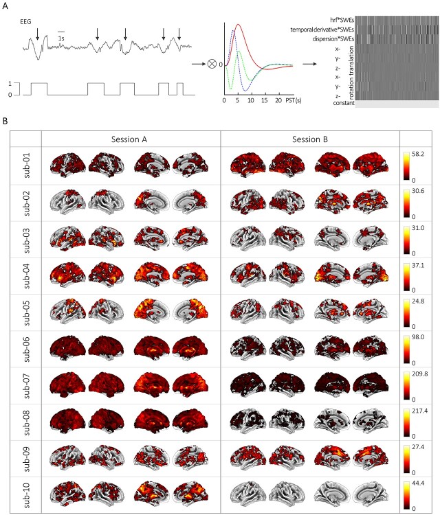
Figure 3.
SWE-related hemodynamic changes at the single-subject, single-session level. (A) Scheme of the fMRI data analysis procedure. From left to right: Individually detected SWEs were converted to SWE vectors including the precise timing of the events. These SWE vectors were convolved with the canonical HRF and its temporal and dispersion derivative for event-related fMRI analyses. An exemplary design matrix shows the individual GLM which contains the SWE vector convolved with the canonical HRF and its derivatives in time and dispersion, six movement parameters, and a constant term. (B) BOLD activation maps showing significant changes upon SWE appearance (voxel-level: clusters >50 voxels at Puncorr < 0.001; cluster-level: PFWEcorr < 0.05) for sessions A and B of all subjects, respectively. Color bars indicate F-values. Of note, intrasubject variability can be appreciated comparing between sessions, for example, sub-04 session A versus sub-04 session B.

