. 2022 Oct 23;82(21):3962–3973. doi: 10.1158/0008-5472.CAN-21-4339

Figure 3.

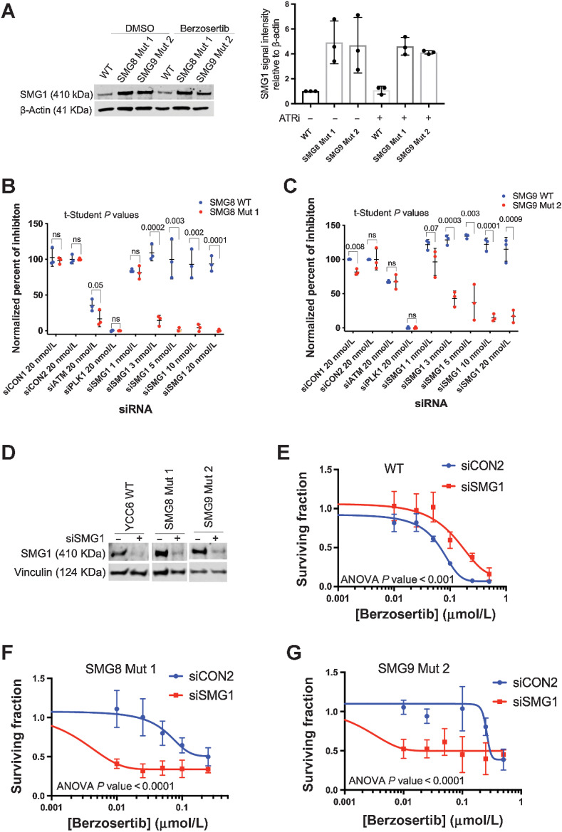
Figure 3. Silencing of SMG1 resensitizes SMG8 and SMG9 knockout cells to ATR inhibition. A, SMG1 is overexpressed in the SMG8 and SMG9 mutant cells after 48 hours of DMSO or 150 nmol/L berzosertib exposure, compared with the WT cells (Left). Smg1 signal intensity relative to β-Actin expression representing relative protein expression using ImageJ (right). Error bars represent SEM, considering three biological replicates. B and C, SMG1 knockdown experiment (384-well plates) showing an increase in the normalized percent of inhibition in the SMG8 and SMG9 mutant cells (red) compared with the WT cells (blue) following exposure to a range of SMG1 siRNA concentrations (from 1 to 20 nmol/L; negative controls; siCON1, siCON2, positive controls; siATM, siPLK1). D, Western blot analysis showing siSMG1 silencing in the YCC6 WT and SMG8 and SMG9 mutant cells. E–G, Silencing of SMG1 in YCC6 WT, SMG8 and SMG9 mutant cells resensitizes them to ATRi (384-well plate, 5-day assay). siCON2 is used as a negative control. All panels of this figure are representative of three or more biological replicates.

Silencing of SMG1 resensitizes SMG8 and SMG9 knockout cells to ATR inhibition. A, Left, SMG1 was overexpressed in the SMG8 and SMG9 mutant cells after 48 hours of DMSO or 150 nmol/L berzosertib exposure compared with the WT cells. Right, Smg1 signal intensity relative to β-actin expression representing relative protein expression using ImageJ. Error bars represent SEM, considering three biological replicates. B and C, SMG1 knockdown experiment (384-well plates) showing an increase in the normalized percent of inhibition in the SMG8 and SMG9 mutant cells (red) compared with the WT cells (blue) following exposure to a range of SMG1 siRNA concentrations (from 1 to 20 nmol/L; negative controls, siCON1, siCON2; positive controls, siATM, siPLK1). D, Western blot analysis showing siSMG1 silencing in the YCC6 WT and SMG8 and SMG9 mutant cells. E–G, Silencing of SMG1 in YCC6 WT, SMG8, and SMG9 mutant cells resensitizes them to ATRi (384-well plate, 5-day assay). siCON2 was used as a negative control. All panels of this figure are representative of three or more biological replicates. ns, nonsignificant.