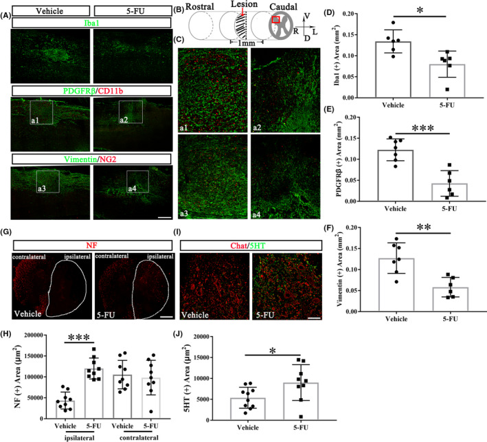
FIGURE 3.

5‐FU administration attenuates fibrotic scar formation. (A) Immunohistochemistry of rat coronal spinal sections at 28dpo. Scale bar = 200 μm. (B) Schematic representation of the lesion and displayed regions (red box) analyzed by Image J to determine fibrotic scar formation (Figure A–F) and axonal regeneration/preservation (Figure 3G–J). (C) The enlargements of a1‐a4 from Figure A. (D–F) Quantification of expression areas of Iba1, PDGFRβ and vimentin (n = 6/group). (G) Immunohistochemistry cross section of rat spinal cord at 28dpo with anti‐NF antibody staining. White line area indicates the ipsilateral spinal cord. Scale bar = 100 μm. (H) Quantification of the NF positive area (n = 9/group). (I) Double immunofluorescence labelling of the spinal cord anterior horn at 28dpo with anti‐Chat and anti‐5HT antibodies. (J) Quantification of the 5HT positive area (n = 9/group). Graphical data are presented as the mean ± standard deviation. Scale bar = 50 μm. DAPI (blue) labeled cell nucleus. R: right, L: left, V: ventral, D: dorsal. *p < 0.05, **p < 0.01, ***p < 0.001
