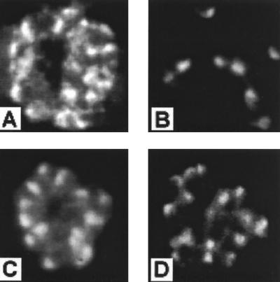
FIG. 2.
Distribution of staining on immunofluorescence analysis of culture supernatants by MAbs 5G2 (A) and 3A9 (B) and of serum from rabbit 2 (diluted 1:1000) (C) and rhesus monkey VH2 (diluted 1:500) (D). Merozoites of P. vivax ONG strain parasites developing within schizont-infected erythrocytes (A, C, and D) and as free merozoites following release from schizonts (B) show the typical apically restricted pattern of staining consistent with AMA-1 localization in rhoptries (24). Identical patterns of distribution were seen for all serum samples from rhesus monkeys immunized with PV66Δglyc43–487.

