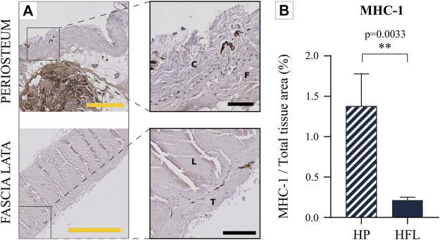
FIGURE 8.
Comparison of the HP (top) and HFL (bottom) by immunohistochemistry for MHC-1 (A) (including zoom in) and MHC-1 quantification (N = 4 donors x three samples from each donor leading to n = 12 specimens analyzed for each tissue) (B). C: cambial layer, F: fibrous layer, L: longitudinal layer, T: transverse layer, HP: human periosteum, HFL: human fascia lata. Black scale bars: 100 µm. Yellow scale bars: 500 µm.

