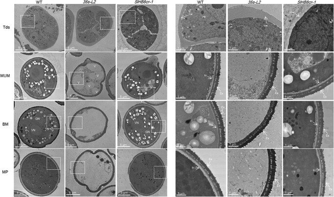
Figure 4.
Transmission electron microscopy of pollen development in the wild-type, SlHB8 gene knockout and p35s::SlHB8Ris lines. BM: binucleate microspore stage; cw: callose wall; Ex: exine; GN: generative nuclei; In: intine; MP: mature pollen stage; MUM: middle uninucleate microspore stage; Tds: tetrad stage; V: vacuole; VN: vegetative nuclei. The scale bar indicated 5 μm (left: whole organ) and 1 μm (right: magnified location), respectively.

