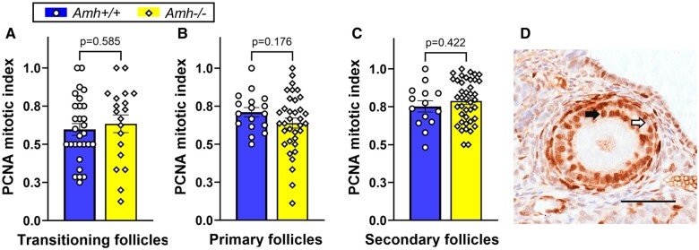
Figure 2.
Granulosa cell mitotic index as a measure of follicle development rate in adult Amh+/+ and Amh−/− mice. (A) transitioning follicles (Amh+/+: n = 29, Amh−/−: n = 19), (B) primary follicle (Amh+/+: n = 17, Amh−/−: n = 35) and (C) secondary follicles (Amh+/+: n = 14, Amh−/−: n = 41) showed no significant difference between the genotypes (Student’s t-test). (D) Example of PCNA immunohistochemical staining (black arrow) and unstained (white arrow) granulosa cells in a secondary follicle from an Amh−/− mouse. Data are expressed as mean ± SEM Scale bar: 50 µm. AMH, anti-Müllerian hormone; PCNA, proliferating cell nuclear antigen.

