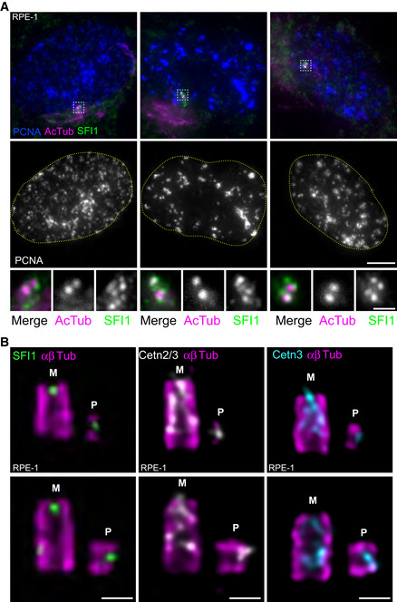
Figure 2. SFI1 and Centrin are recruited at the onset of centriole biogenesis.

-
ARepresentative confocal images of RPE‐1 cycling cells stained for SFI1 (green), acetylated tubulin (AcTub, magenta), and PCNA (blue). DNA boundaries are marked with a yellow dotted line. White dashed line squares correspond to insets. Scale bar: 5 μm.
-
BRepresentative confocal images of expanded duplicating centrioles from RPE‐1 cells stained for α/β‐tubulin (αβTub, magenta) and SFI1 (green, left panel), Centrin 2/3 (Cetn2/3, gray, middle panel) or Centrin 3 (Cetn3, cyan, right panel). M stands for mature centriole and P stands for procentriole. Note that both SFI1 and Centrins are recruited very early at procentrioles as a distal dot. Scale bars: 200 nm.
