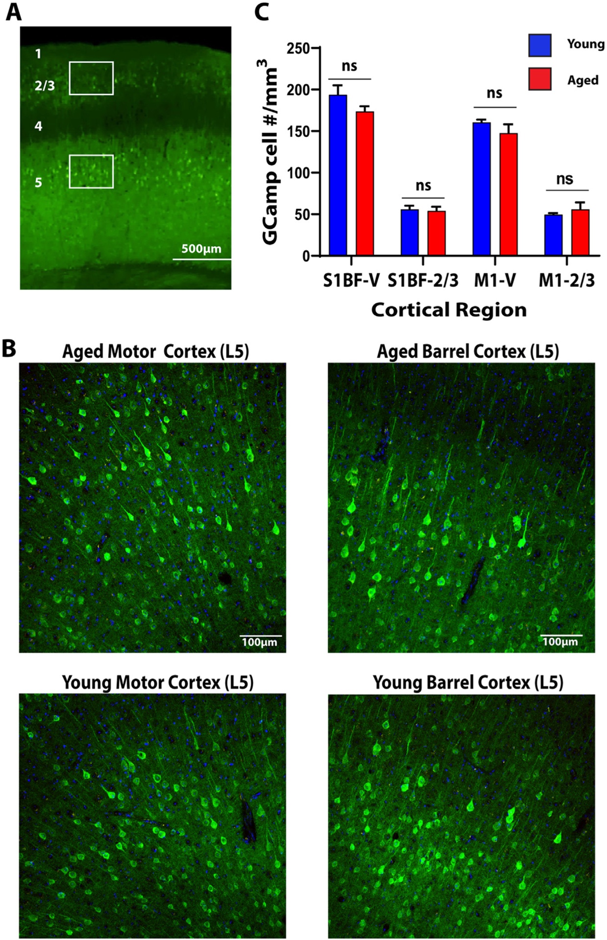
Fig. 1. Young and Aged Mice have Similar Cortical GCaMP Expression.

A. Low magnification view of the cortex. Numbers represent cortical layers. Boxes represent regions of interest in which counts were undertaken. B. Representative expression patterns from layer 5 motor and barrel cortex in aged and young animals. C. Quantification of GCaMP positive cell count in Layer 2/3 and Layer 5 barrel and motor cortices in aged (red) vs young (blue) animals.
