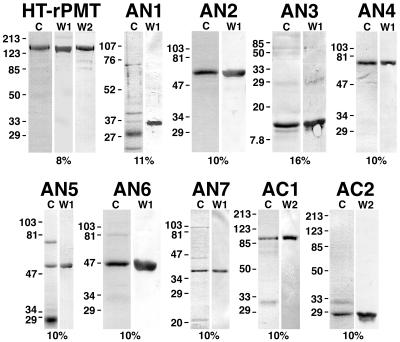
FIG. 2.
Coomassie-stained SDS-polyacrylamide gel and Western blot analysis of Ni2+ chelate affinity-purified samples of rPMT and recombinant PMT fragments used in this study. The toxin protein sample analyzed is indicated above each set of gels. Lanes: C Coomassie-stained SDS-polyacrylamide gels; W1 and W2, the corresponding Western blots with anti-toxA28–42 and anti-toxA1239–1253, respectively, as primary antibodies. The percentage of acrylamide used for the gel is indicated below each set of gels. In each lane, 3 to 5 μg of protein was loaded. The positions of molecular weight markers are indicated in kilodaltons at the left. HT-rPMT denotes rPMT before removal of the His tag. The ToxAN2 and ToxAN3 samples shown were after removal of the His tag, and the ToxAN1 sample shown was before removal of the His tag.

