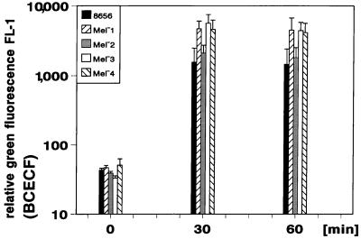
FIG. 3.
Kinetics of phagocytosis of E. dermatitidis as determined by flow cytometry. The subsequent increase of relative green fluorescence of neutrophils after association with the respective BCECF-labeled E. dermatitidis strains is shown. The mean values ± SD of six independent assays are displayed.

