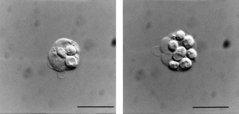
FIG. 4.
Intracellular location of the BCECF/AM-stained E. dermatitidis 8656 as determined by interference contrast microscopy. After 60 min of incubation in heparinized blood, the yeast cells are located within the neutrophils (Bar = 10 μm). (Left panel) Neutrophil with two yeast cells ingested, one of them budding. (Right panel) Neutrophil with maximum number of yeast cells ingested. These microorganisms could be also identified as yeast cells due to their green fluorescence in simultaneously performed epifluorescence microscopy studies.

