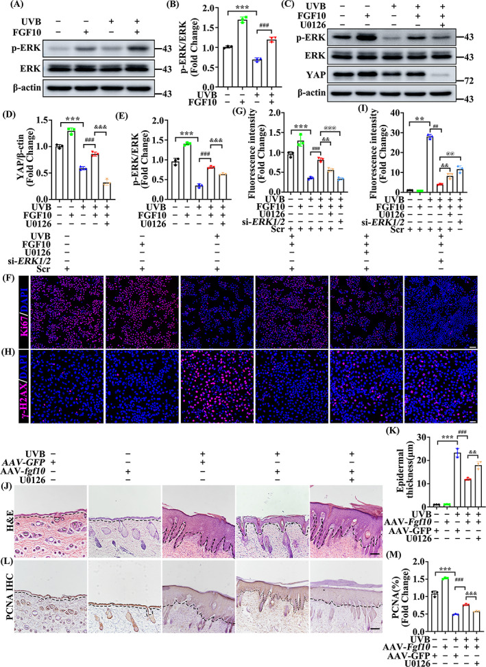
FIGURE 6.

FGF10 protects UVB‐induced skin injury through the ERK‐YAP pathway. (A) The expression levels of p‐ERK and ERK in the untreated or UVB treated with or without FGF10. β‐Actin was used as a loading control (n = 3). (B) Quantification of p‐ERK/ERK levels in (A). (C) The protein expression of p‐ERK, ERK and YAP were measured by immunoblotting assay in untreated or UVB treated with or without FGF10 in the presence or absence of U0126. β‐Actin was used as a loading control (n = 3). (D,E) Quantification of YAP, p‐ERK/ERK levels in (C). (F) Immunofluorescence staining of Ki‐67 (Red) in the untreated or UVB treated HaCaT with or without FGF10 in the presence or absence of U0126 and si‐ERK1/2. Nuclei were stained with DAPI (Blue) (n = 3). Scale bars = 100 μm. (G) Quantification the percentage of Ki67‐positive cells from (F). (H) Immunofluorescence staining of γ‐H2AX (Red) in the untreated or UVB treated HaCaT with or without FGF10 in the presence or absence of U0126 and si‐ERK1/2. Nuclei were stained with DAPI (Blue) (n = 3). Scale bars = 50 μm. (I) Quantification the percentage of γ‐H2AX‐positive cells from (H). (J) Representative HE staining of AAV‐GFP or AAV‐Fgf10 transfected skin treated with or without UVB in the presence or absence of U0126 (n = 3). Scale bars = 100 μm. (K) Quantification the epidermal thickness from (J). (L) Immunohistochemical staining of PCNA in the AAV‐GFP or without UVB and AAV‐Fgf10 transfected skin treated with or without UVB in the presence or absence of U0126. The positive staining (brown) demonstrated positive expression (n = 3). Scale bars = 100 μm. (M) Quantification the PNCA‐positive from (L). The data are presented as the means ± SEM. ***P < 0.001, **P < 0.01, ### P < 0.001, ## P < 0.01, &&& P < 0.001, && P < 0.01, @@@ P < 0.001, @@ P < 0.01 vs. the corresponding untreated group
