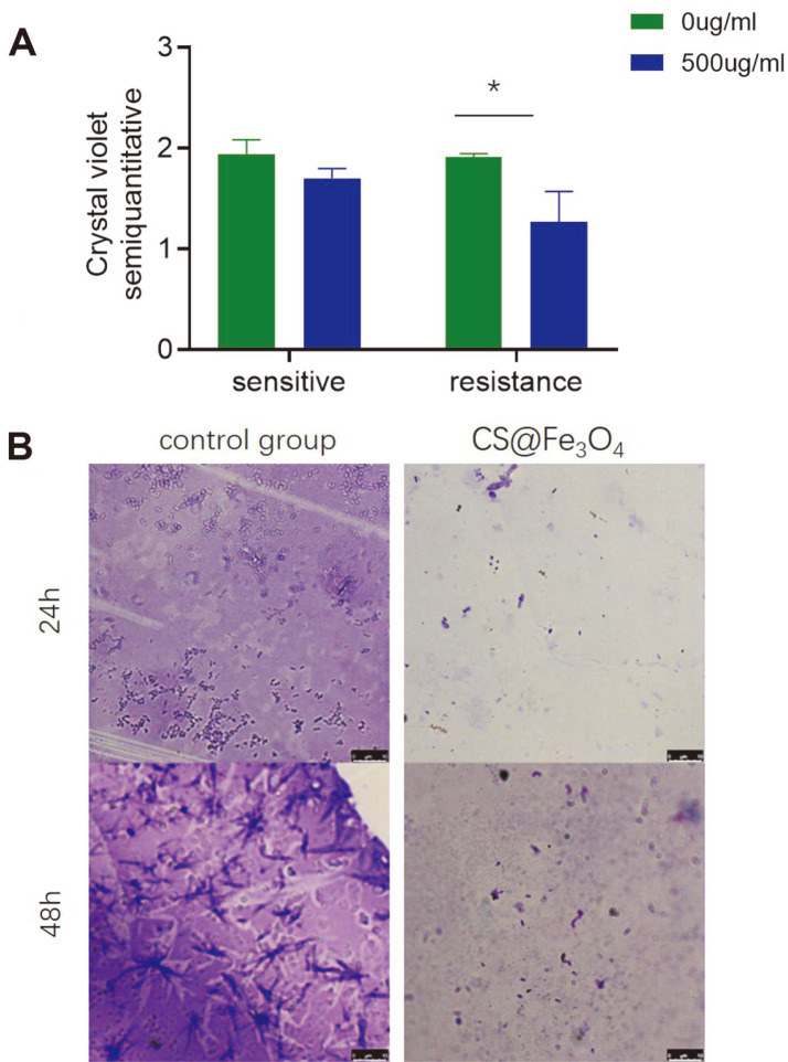
Fig. 2. A. The semi-quantitative crystal violet staining assay was used to determine the amount of bacterial biofilm.
OD values are given as the mean ± SD of three independent experiments. * p < 0.05. B. Microscopic examination of the effects of CS@Fe3O4 nanoparticles on biofilm formation were observed by crystal violet staining after 24 and 48 h.

