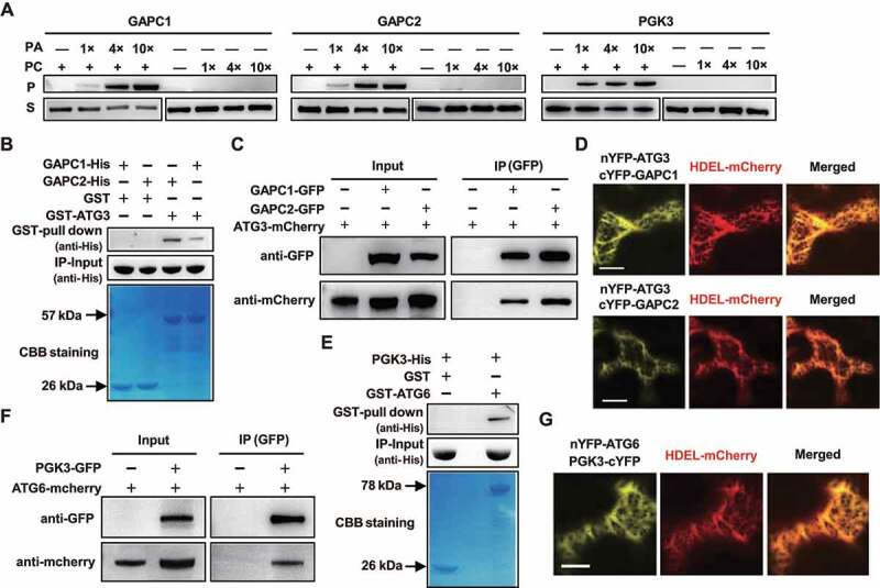
Figure 3.

GAPCs interacted with ATG3, and PGK3 interacted with ATG6, at endoplasmic reticulum (ER). (A) Liposome binding assay showed the correlation between bound GAPCs or PGK3 and PA. Purified His-tagged GAPCs or PGK3 were incubated with liposomes containing di18:1-PC only or di18:1-PA/PC (1/3 mole ratio). 1×, 4×, and 10× refer to the concentration gradient of PC or PA/PC liposomes used. Bound (pellet with liposomes, P) GAPCs or PGK3 and non-binding protein (supernatant with liposomes, S) were detected by immunoblotting. (B) GST pull-down assays revealed that GAPC1-His and GAPC2-His directly interact with GST-ATG3. Recombinantly expressed (from E. coli) GST or GST-ATG3 proteins were incubated with GAPC1-His or GAPC2-His and analyzed by immunoblot assays using an anti-His antibody (upper panel). Input of His-tagged proteins (middle panel) and equal aliquots of glutathione beads loaded with GST-ATG3 or GST (separated by SDS-PAGE and stained with Coomassie blue, bottom panel) were shown. (C) Co-IP assay demonstrated the interaction between ATG3-mCherry and GAPC1-GFP or GAPC2-GFP in vivo. GFP-tagged GAPCs was transiently co-expressed with mCherry-tagged ATG3 in N. benthamiana leaves and immunoprecipitated by GFP affinity magnetic beads. Resultant immunoprecipitation (IP) and cell lysate were analyzed by immunoblotting using anti-GFP or anti-mCherry antibodies. (D) Fluorescence observation of N. benthamiana leaf epidermal cells co-expressing GAPC1-cYFP/GAPC2-cYFP and nYFP-ATG3 with ER marker HDEL-mCherry. Representative images were shown (bar = 50 μm). (E) GST pull-down assays revealed that PGK3-His directly interacts with GST-ATG6. Recombinantly expressed (from E. coli) GST or GST-ATG6 proteins were incubated with PGK3-His and analyzed by immunoblot assays using an anti-His antibody (upper panel). Input of His-tagged proteins (middle panel) and equal aliquots of glutathione beads loaded with GST-ATG6 or GST (separated by SDS-PAGE and stained with Coomassie blue, bottom panel) were shown. (F) Co-IP assay demonstrated the interaction between ATG6-mCherry and PGK3-GFP in vivo. GFP-tagged PGK3 was transiently co-expressed with mCherry-tagged ATG6 in N. benthamiana leaves and immunoprecipitated by GFP affinity magnetic beads. Resultant immunoprecipitation (IP) and cell lysate were analyzed by immunoblotting using anti-GFP or anti-mCherry antibodies. (G) Fluorescence observation of N. benthamiana leaf epidermal cells co-expressing PGK3-cYFP and nYFP-ATG6 with ER marker HDEL-mCherry. Representative images were shown (bar = 50 μm).
