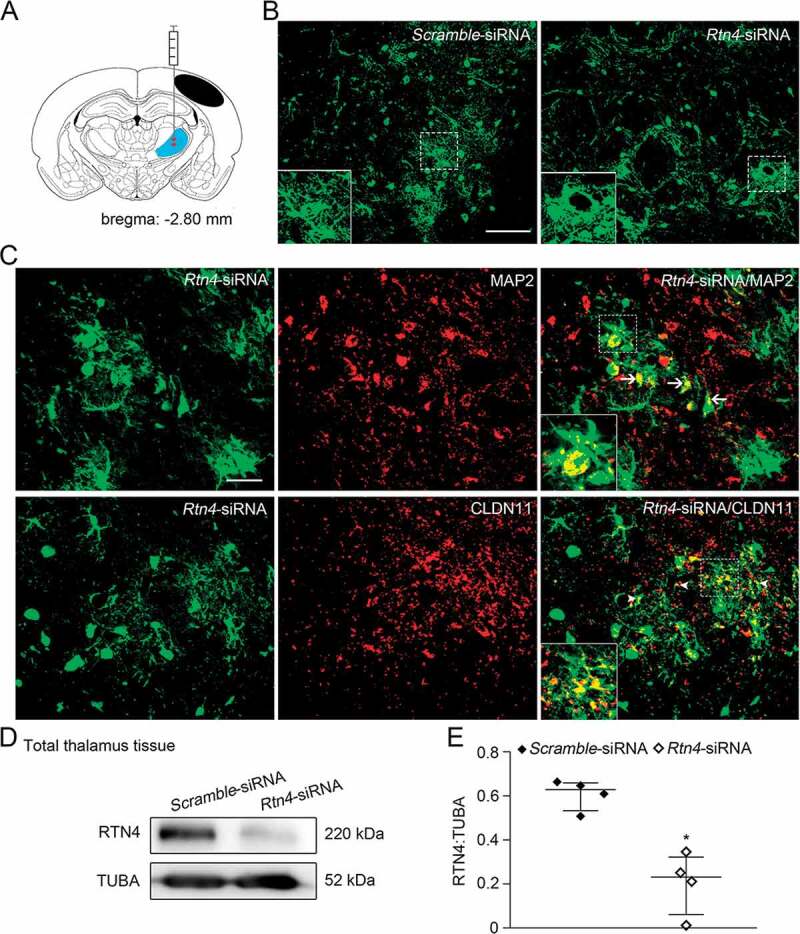
Figure 4.

Knockdown of Rtn4 mediated by siRNA in the ipsilateral thalamus after cortical infarction. (A) Schematic diagram of brain section (−2.80 mm from bregma) shows the location of cortical infarction (black area) and the ipsilateral thalamus (blue area) where siRNA-lentivirus is delivered to four sites (red). (B) GFP-tagged Scramble- and Rtn4-siRNA were detected in the ipsilateral thalamus at 7 days after MCAO. Scale bar: 100 μm. (C) Immunostaining shows GFP-tagged Rtn4-siRNA to be expressed predominantly on MAP2+ neurons (arrows) and CLDN11+ oligodendrocytes (arrowheads). Scale bar: 50 μm. (D) Immunoblotting shows RTN4 expression in the ipsilateral thalamus from the Scramble- and Rtn4-siRNA groups at 7 days after MCAO. (E) Quantitative analysis of RTN4 level relative to TUBA. n = 4, data are expressed as median ± interquartile range. *P < 0.05, compared with the Scramble-siRNA group.
