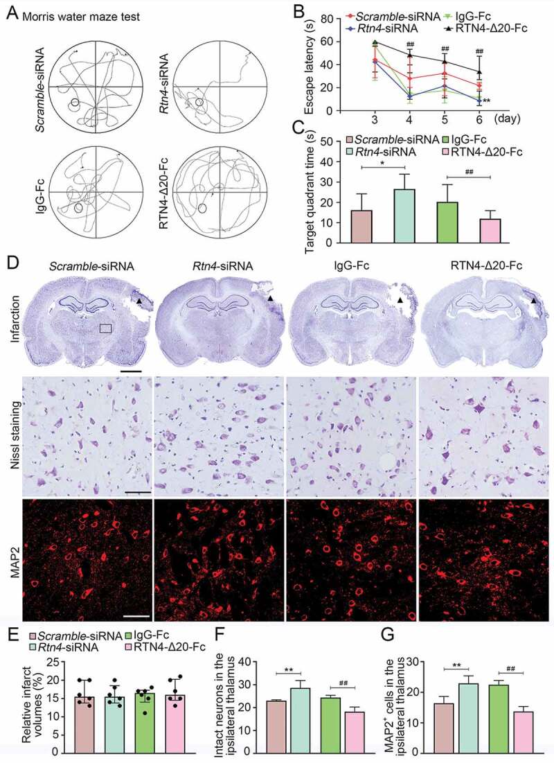
Figure 10.

Cognitive function and secondary neuronal damage of the ipsilateral thalamus after cortical infarction. (A) The representative swimming path of the animals in the Scramble-siRNA, Rtn4-siRNA, IgG-Fc, and RTN4-Δ20-Fc groups at 7 days after MCAO. (B) Animals in the Rtn4-siRNA group showed a shorter escape latency to reach the platform on day 6 after MCAO than that in the Scramble-siRNA group. In contrast, animals treated with RTN4-Δ20-Fc had longer escape latency from day 4–6 after MCAO than those in the IgG-Fc group. **P < 0.01, compared with the Scramble-siRNA group, ##P < 0.01, compared with the IgG-Fc group. (C) On day 7 after MCAO, animals of the Rtn4-siRNA group spent longer target quadrant time than that in the Scramble-siRNA group while animals treated with RTN4-Δ20-Fc showed less time in the target quadrant compared to the IgG-Fc group. n = 8, data are expressed as median ± interquartile range. *P < 0.05, compared with the Scramble-siRNA group, ##P < 0.01, compared with the IgG-Fc group. (D) Representative Nissl staining indicates the cortical infarction (black triangle) and neuronal damage within the ipsilateral thalamus (black rectangle) of the Scramble-siRNA, Rtn4-siRNA, IgG-Fc, and RTN4-Δ20-Fc groups. Immunostaining analysis of MAP2+ neurons within the ipsilateral thalamus across the different groups. Scale bar: 2 mm, 50 μm. (E-G) Quantitative analysis of the relative infarct volumes, and the number of intact neurons and MAP2+ cells. n = 6, data are expressed as median ± interquartile range. **P < 0.01, compared with the Scramble-siRNA group, ##P < 0.01, compared with the IgG-Fc group.
