Figure 8.

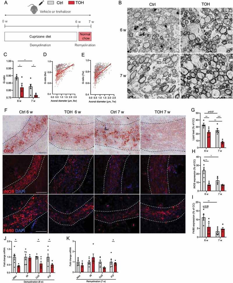
Trehalose stimulates remyelination in the cuprizone model. (A) Experimental setup of cuprizone-induced demyelination in vivo model. (B) Representative images of transmission electron microscopy analysis of corpus callosum (cc) from vehicle (Ctrl)- and trehalose (TOH)-treated animals after demyelination (6 w) and during remyelination (7 w). Scale bar: 100 µm. (C-E) Analysis of the g-ratio (the ratio of the inner axonal diameter to the total outer diameter) and g-ratio as a function of axon diameter in cc. (F) Representative images of Oil Red O (ORO) staining (scale bar: 50 µm), immunofluorescence NOS2 staining (scale bar: 100 µm), and immunofluorescence ADGRE1 staining (scale bar: 100 µm). (G) Quantification of ORO staining (lipid load defined as percent area covered in lipid droplets of the total cc area). (H,I) Relative NOS2+ area and ADGRE1+ area of cc. (J,K) mRNA expression of inflammatory mediators in CC. n = 4–7. All data are represented as mean ± SEM. *p < 0.05, **p < 0.01, and ***p < 0.001.
