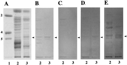
FIG. 3.

Binding of anti-blood group MAb to neutral glycosphingolipids. Neutral glycosphingolipids (100 μg) from phenotypes A (lane 2) and D (lane 3) pigs were separated on HPTLC plates as described in Materials and Methods. Chromatograms were stained with orcinol-sulfuric acid reagent (A) or incubated with the anti-Lex (B), anti-H (C), anti-B (D), and anti-A (E) MAb as described in Materials and Methods. Lane 1 contains glycolipid standards Lc2Cer (2), Gb4Cer (4) and Gb5Cer (5). The arrowheads indicate the positions of IGLad.
