Fig. 3.
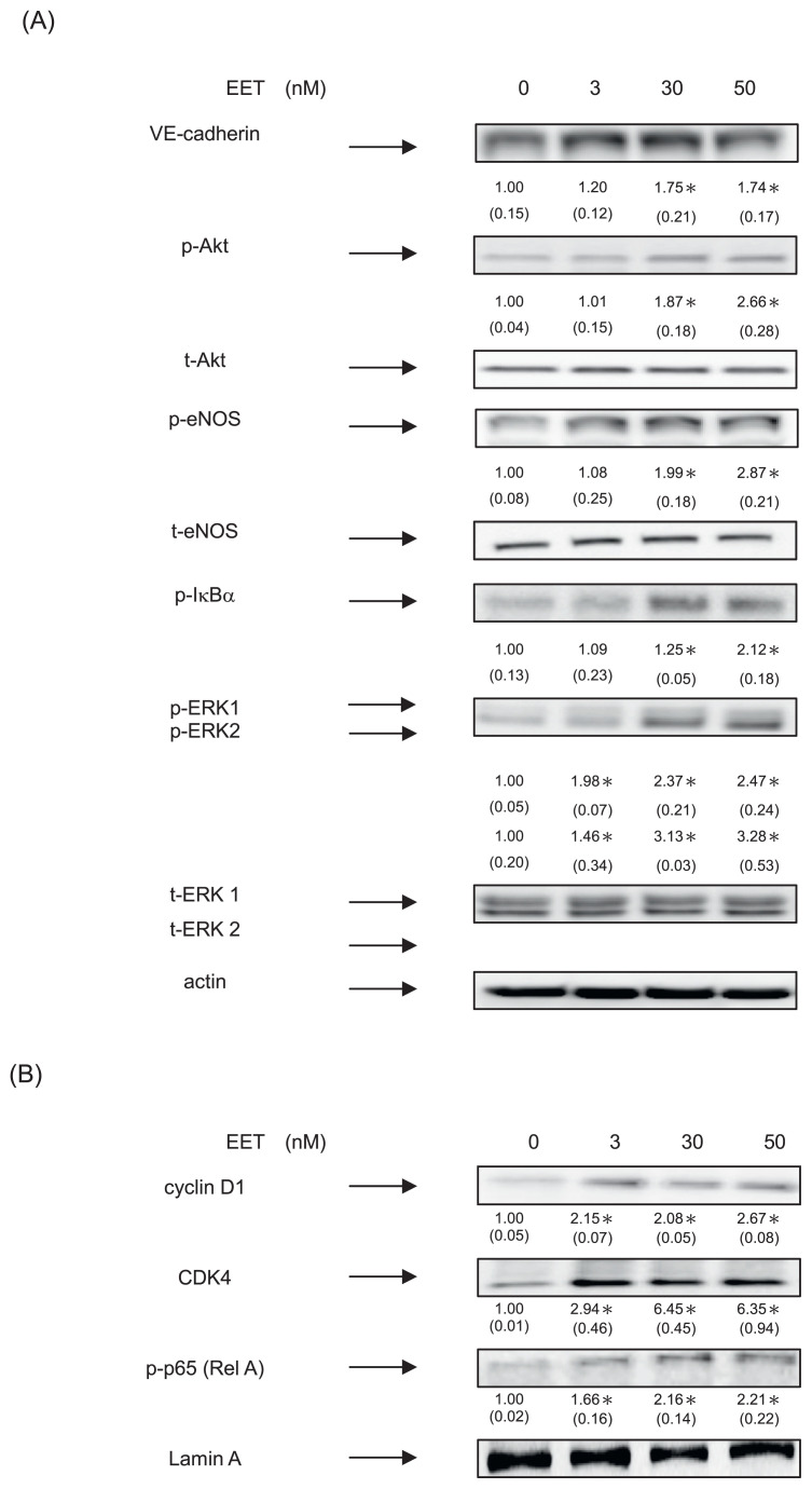
11,12-EET induced neovasculogenesis through increment of phosphorylated Akt, eNOS and ERK 1/2 proteins in hEPCs. hEPCs were treated with 11,12-EET (at concentrations of 0, 3, 30 and 50 nM) for 8 h. (A) Measurement of cytoplasmic proteins including VE-cadherin, p-Akt, t-Akt, p-eNOS, t-eNOS, p-IκBα, p-ERK1/2, t-ERK 1/2 and actin was performed by using Western Blotting analysis as described in Materials and Methods. The integrated densities (mean ± SD) of each protein (VE-cadherin, p-IκBα, p-Akt, p-eNOS, p-ERK1/2) are adjusted with the corresponding control proteins (actin, t-Akt, t-eNOS or t-ERK 1/2) and shown in the bottom row. A single asterisk indicates a statistical difference in comparison with the 11,12-EET untreated control group (P < 0.05). (B) Analysis of nuclear proteins were conducted using antibodies against cyclin D1, CDK4, p-p65 (RelA) and lamin A. The integrated densities (mean ± SD) of these proteins are adjusted with the loading control lamin A protein are shown in the bottom row. A single asterisk represented a statistical difference in comparison with 11,12-EET-untreated control group (P < 0.05).

