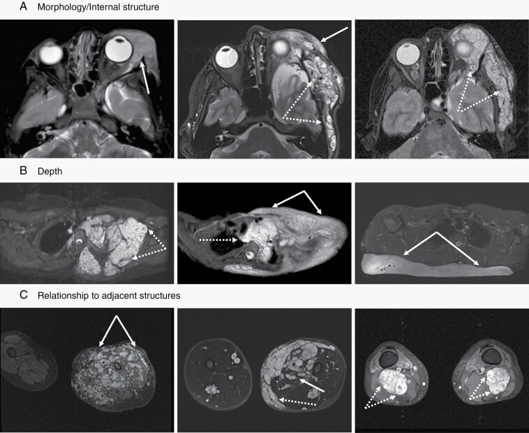
Fig. 2.
MRI examples of plexiform neurofibromas (PN) demonstrating tumor characteristics used in the proposed classification schema. All images are fat suppressed with STIR (short tau inversion recovery) technique. (A) The internal structure can be homogeneous without notable architectural elements (left panel, solid arrow), appear as conglomerate of small nodules (right panel, dotted arrows), or show a combination of both features (middle panel). (B) Any portion of peripheral nerves may be affected by PN. Proximal nerve segments give rise to deep internal PN (left panel, dotted arrows), superficial PN (right panel, solid arrows) are associated with terminal nerve branches, but many lesions have components of both (middle panel). (C) The interface between PN and surrounding tissues can range from interdigitating and intricately connected (left panel, solid arrows) to sharply defined and well separated (right panel, dotted arrows), with most lesions falling in between those two extremes (middle panel).

