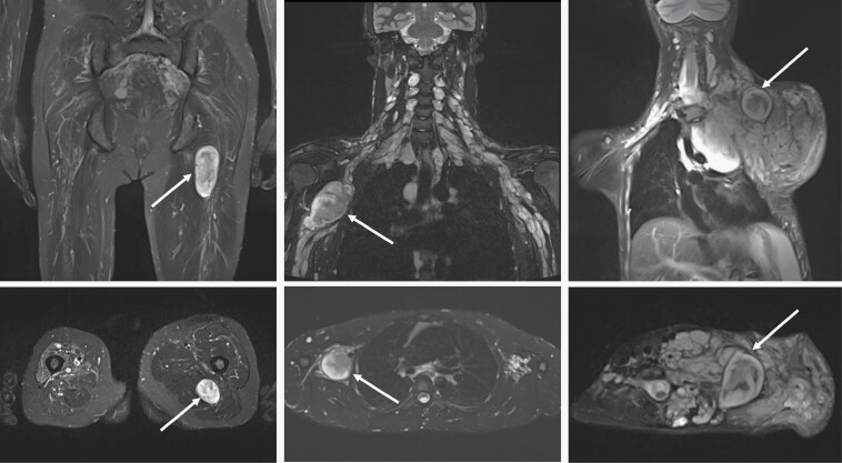
Fig. 3.
Coronal (top row) and axial (bottom row) STIR MRI examples of distinct nodular lesions (DNL) in patients with NF1. The left panels show a DNL (arrow) arising from the left sciatic nerve. In the middle panels, plexiform neurofibroma (PN) can be seen along the brachial plexus on both sides, with a prominent nodule present on the right (arrow). On the right, the DNL (arrow) stands out from the background of a large neck, shoulder, and chest PN.

