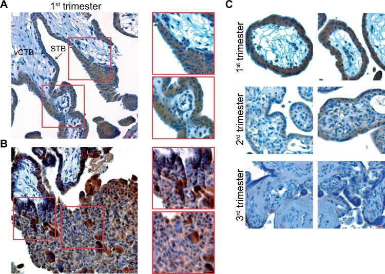
Figure 1.
BCL6 is expressed in various trophoblast types of first trimester placentas and its expression declines during gestation. (A) First trimester placental tissue sections were immunohistochemically stained for BCL6 and DNA. Left: a representative image is shown. Scale: 50 µm. Right: enlarged villi. Scale: 20 µm. STB, syncytiotrophoblast; vCTB, villous cytotrophoblast. (B) Representative EVTs positively stained with BCL6 antibody in the decidua of first trimester placenta (left, scale: 50 µm) and enlarged regions with positive EVTs (right, scale: 20 µm). EVT, extravillous trophoblast. (C) Representative images are shown for first (upper panel), second (middle panel) and third trimester placental tissues (lower panel) stained for BCL6 and DNA. Scale: 50 µm.

