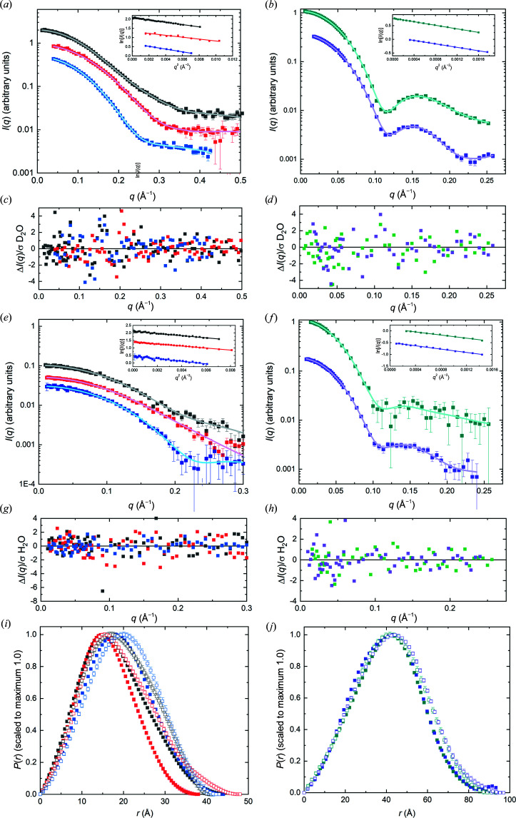
Figure 5.
(a, b) I(q) versus q (symbols) with P(r) model fits (black lines) from consensus SANS data in D2O for each protein, with Guinier plots (to qR g = 1.3) as insets. (c, d) Error-weighted residual plots for the fits in (a) and (b), respectively. (e) and (f) are the same plots as in (a) and (b) but for SANS data in H2O, with (g) and (h) showing the corresponding error-weighted residual plots. (i, j) Corresponding P(r) versus r plots for the fits in (a) and (b) (D2O, solid squares) and (e) and (f) (H2O, open squares). The colour key throughout is RNaseA, black; lysozyme, red; xylanase, blue; urate oxidase, dark cyan; xylose isomerase, purple. Error bars are standard errors, and where not apparent are smaller than the symbols.

