
FIG. 1.
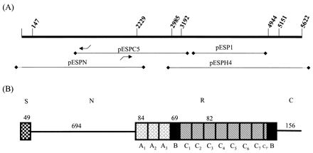
Schematic of the esp gene and inferred protein product. (A) Structural esp gene and location of insert DNA from various clones used to derive the complete sequence of esp. Bent arrows indicate the positions of oligonucleotide primers esp15 and esp16 used for inverse PCR. (B) Deduced Esp protein showing the inferred signal (S), N-terminal (N), repeat (R), and C-terminal (C) regions. Numbers above each region denote the number of amino acid residues in each segment. The repeat blocks A, B, and C are identified, with C7, denoting the partial C repeat.