. 2022 Oct 12;11:e80165. doi: 10.7554/eLife.80165

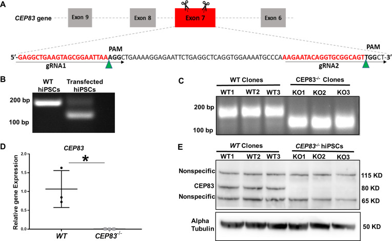
Figure 1. Generation of centrosomal protein 83 (CEP83)-deficient human pluripotent stem cells.

(A) Schematic of the experimental approach to induce a deleting mutation in exon 7 of the CEP83 gene (as described in the methods section). (B) DNA extracted from pooled transfected cells was subjected to PCR, targeting the predicted deletion site in the CEP83 gene. In addition to the 182 bp fragment present in untransfected wildtype (WT) cells, an approximately 120 bp fragment was detected in transfected cells, corresponding to the induced deletion in exon 7. (C) Three clones (CEP83−/− clones KO1, KO2, KO3) carried 62–74 bp deletions within CEP83 exon 7, which led to an induction of premature stop codons or frameshift mutation on both alleles of CEP83. Three wildtype clones (WT1, WT2, and WT3) were used as controls. (D) Quantitative RT-PCR for a fragment corresponding to the deleted region in CEP83 exon 7 produced a detectable signal in RNA extracts from WT clones but not CEP83−/− clones. (E) Immunoblotting of WT and CEP83−/− clones using a CEP83 antibody targeting the C-terminal region of the protein (see Methods for details) indicated a complete loss of the 83 KD band corresponding to CEP83 protein in the three KO clones compared with the three WT clones. Data are mean ± SD.*p<0.05 and **p<0.01 vs. WT. See Figure 1—source data 1 and 2. See also Figure 1—figure supplements 12.

Figure 1—source data 1. The file contains detailed original PCR gels and immunoblots.
Figure 1—source data 2. Excel sheet shows RT-qPCR data for mRNA expression of CEP83 in WT and knockout hiPSCs.
Figure 1—source data 3. File contains uncropped PCR gels and immunoblots.

Figure 1.

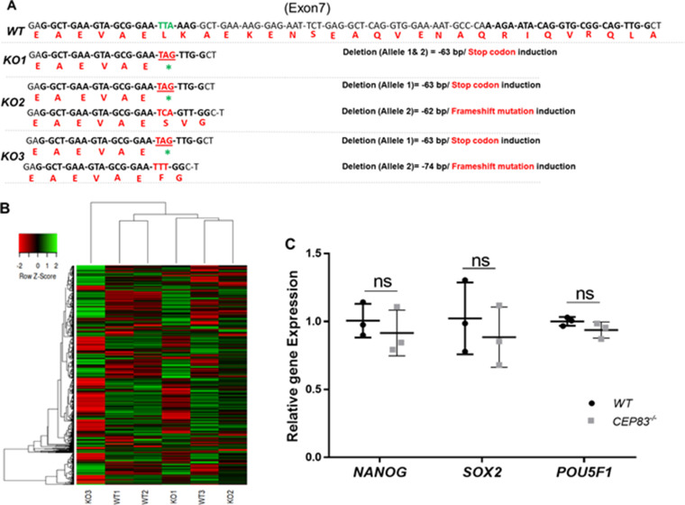
Figure 1—figure supplement 1. CEP83−/− human-induced pluripotent stem cells (hiPSCs) retain global iPCS cell gene expression signatures and express pluripotency markers.

Figure 1—figure supplement 1.

(A) Alignment of the modified KO clones mRNA and expected amino acid sequences with WT revealed induction of stop codon on both strands of KO1 clone. While KO2 clone shows induction of stop codon on one allele and frameshift mutation within the second allele with 62 bp deletion. KO3 clone sequence shows induction of stop codon on one allele and frameshift mutation with 74 bp deletion in the second allele. (B) Heatmap showing the expression of the top 1000 highly variable genes (see Methods, with a selection of transcripts per million ≥10) within WT (WT1, WT2, and WT3) and CEP83−/− hiPSCs (KO1, KO2, and KO3) clones. Unbiased hierarchical clustering of clones indicates that gene expression similarity is not driven by WT or KO status. (C) RT-PCR shows no significant differences in the expression of pluripotency markers NANOG, SOX2, and POU5F1 between WT and CEP83−/− hiPSCs. n=3 hiPSCs clones per group. Data are mean ± SD. ns, not significant.
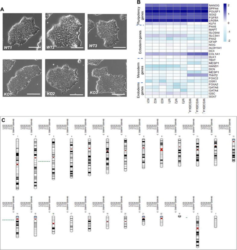
Figure 1—figure supplement 2. Phenotypical, molecular, and genetic characterization of CEP83−/− human-induced pluripotent stem cells (hiPSCs) versus the wildtype hiPSCs.

Figure 1—figure supplement 2.

(A) CEP83−/− hiPSCs clones (KO1, KO2, and KO3) show similar morphology to the WT clones (WT1, WT2, and WT3) under the bright field microscope, scale bar = 200 µm. (B) Using bulk RNA sequencing data, transcripts per million values for marker genes for pluripotency, ectodermal, mesodermal, and endodermal cells were plotted across the samples (KO1, KO2, KO3, WT1, WT2, and WT3). In addition, gene expression of the six samples was compared to three wildtype hiPSCs (WISCi004-A, also referred to as IMR90-4 iPS derived from female lung fbroblasts) that were previously published (Hariharan et al., 2019). (C) The three WT clones, three KO clones, and the parental population were karyotyped using single nucleotide polymorphism - analysis, demonstrating unaffected integrity of karyotypes. Two aberrations (one gain on Chr3 and one gain on Chr14) present in BIHi005-A were previously reported (https://hpscreg.eu/cell-line/BIHi005-A, Berlin Institute of Health Stem Cell Core Facility).