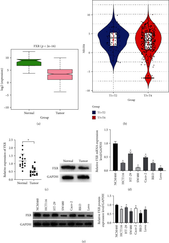
Figure 1.

FXR is lowly expressed in colon cancer cell lines. (a). According to TCGA database, the expression of FXR in colon cancer tissues was significantly reduced compared to normal tissues. The green box shows the normal sample and the pink box shows the tumor sample. (b). According to TCGA database, the expression of FXR in T3+T4 colon cancer patients was significantly reduced compared to T1+T2 patients. The blue violin shows the T1+T2 patients, and the red violin shows the T3+T4 patients. (c). qRT-PCR and Western blot were employed to evaluate the mRNA and protein levels of FXR in colon cancer patients. (d and e). qRT-PCR and Western blot were employed to evaluate the mRNA and protein levels of FXR in colon cancer cell lines. (∗ denotes P < 0.05).
