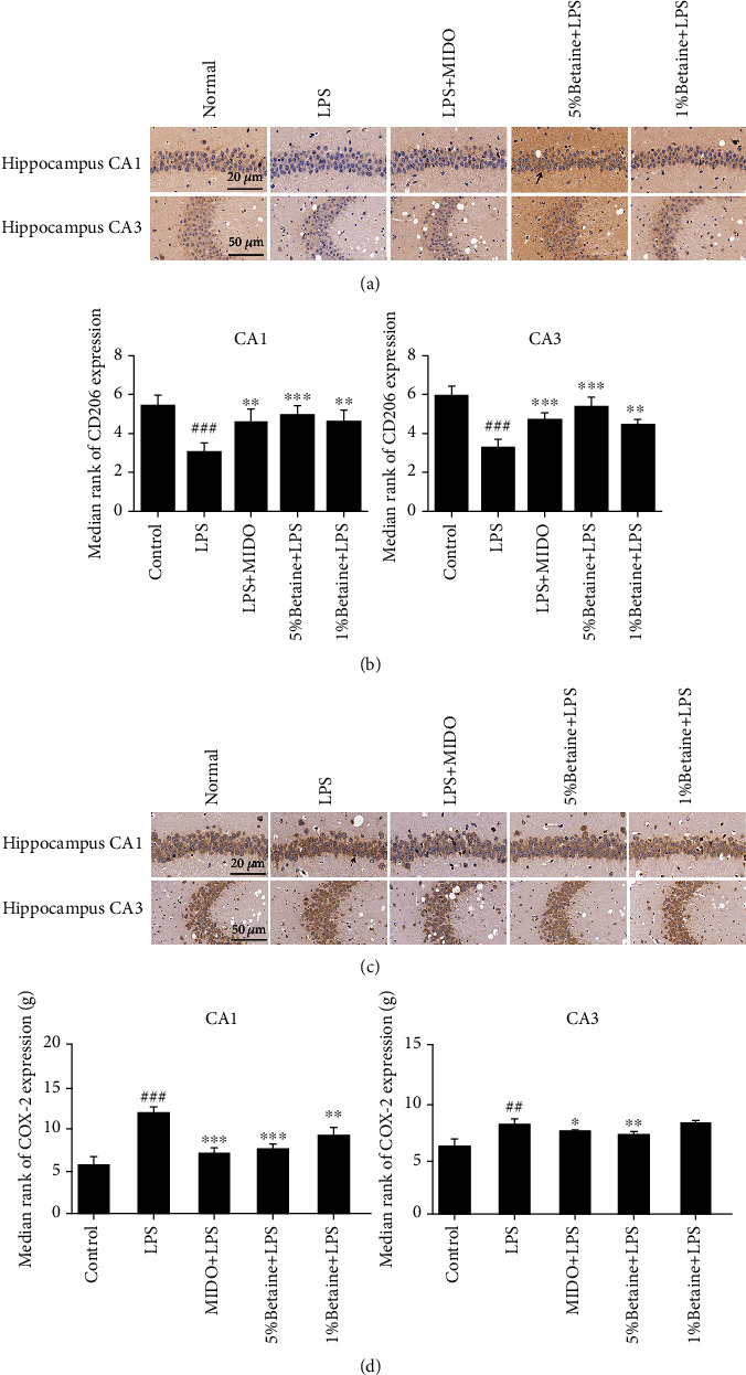
Figure 6.

Immunohistochemical labeling of M1 polarization marker COX-2 and M2 polarization marker CD206 in mice CA1 and CA3 brain regions. Black arrows indicate positive immunohistochemical staining for M1/M2 polarization markers. (a) Typical immunohistochemical results for the effects of betaine on CD206 positive cells in mouse hippocampus. (b) Statistics on the number of CD206 positive cells. (c) Typical immunohistochemical results for the effects of betaine on COX-2 positive cells in mouse hippocampus. (d) Statistics on the number of COX-2 positive cells. The results were expressed as mean ± SEM; ##P < 0.01 and ###P < 0.001 compared to the normal control group; ∗P < 0.05, ∗∗P < 0.01, and ∗∗∗P < 0.001 compared to the model group, n = 6.
