Figure 1.

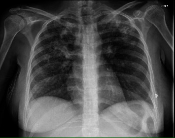
Chest X-ray image showing both alveolar and interstitial infiltrates with findings most prominent within the bilateral upper lobes and right perihilar region.

Chest X-ray image showing both alveolar and interstitial infiltrates with findings most prominent within the bilateral upper lobes and right perihilar region.