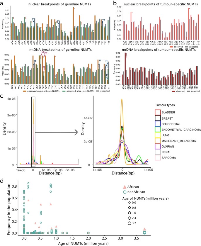
Extended Data Fig. 8. NUMT nuclear breakpoints, relation to PRDM9 binding sites, and NUMT age.
a. Frequencies of trinucleotides around germline NUMTs breakpoints. The breakpoints of nuclear genome are shown at the top and mtDNA genomes at the bottom, common&rare, ultra-rare NUMTs and the expected frequencies shown in the different colours. Trinucleotides of breakpoint flanks more likely occurred in nCC/CCn on mtDNA genome and less likely in nTT/TT on both nuclear and mtDNA genomes, particularly for ultra-rare NUMTs. The same trend was not seen in the tumour-specific NUMTs (b), indicating the signal is driven by biology, but not the sequencing artefacts. b. Frequencies of trinucleotides around tumour-specific NUMTs breakpoints in the nuclear genome (top) and mtDNA genomes (bottom), tumour-specific NUMTs and the expected frequencies shown in the different colours. P values # < 0.1, * < 0.05, < 0.01 **, < 0.001 ***, < 0.0001 **** (two-sided Fisher’s exact test) (Supplementary Table 6). c. Distribution of the distance between PRDM9 binding sites and tumour-specific NUMTs within each tumour type. d. Age of NUMTs estimated in this study. Y axis shows the frequencies of NUMTs in African and non-African populations. The frequencies of NUMTs were different between African and non-African, particularly for the older NUMTs which were more common seen in African population.

