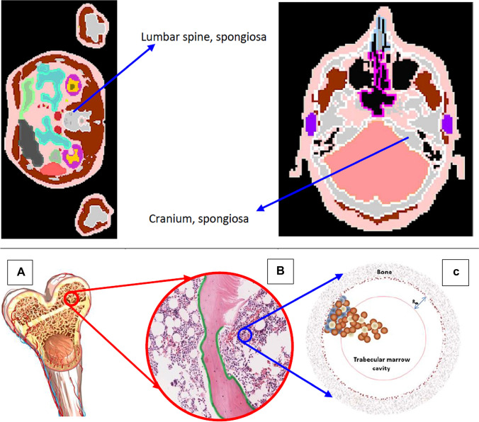
Fig. 1.
Upper panel: ICRP voxel phantom of spongiosa in lumbar spine and cranium (ICRP 2009). Lower panel: from bone to bone marrow phantom (Hobbs et al. 2012), A Structure of upper arm bone (humeri) which shows the spongiosa, the medullary cavity and the cortical bone; B Spongiosa which shows the trabecular bone, red bone marrow, yellow bone marrow and the endosteum; C Mathematical phantom model for red bone marrow, which shows the trabecular marrow cavity and the osteoprogenitor cells (blue), hematopoietic stem and progenitor cells (brown), and adipose cells (white). Figures reproduced with permission by ICRP and IOP Publishing

