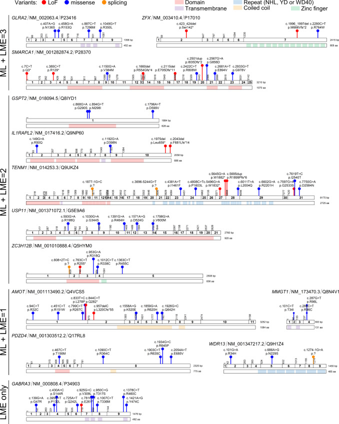
Fig. 7. Damaging variants in selected predicted disorder-associated genes.
Schematic representation of the coding exons, protein domains (when present) and available damaging variants (truncating or CADD ≥ 25) for each selected predicted gene. Types of variants are shown in different colors: lof-of-function (LoF, red), missense (blue), splicing (orange). Protein functional domains are shown: domains (light red), transmembrane segments (purple), NHL, YD or WD40 repeats (light blue), coiled-coils (yellow), zinc-fingers (green). HGVS cDNA and HGVS protein descriptions are shown. The corresponding RefSeq identifier of the MANE Select transcript and Uniprot identifier are shown for each gene. Details of variants displayed in this figure appear in Supplementary Data 10. The schemes were generated with ggplot2.

