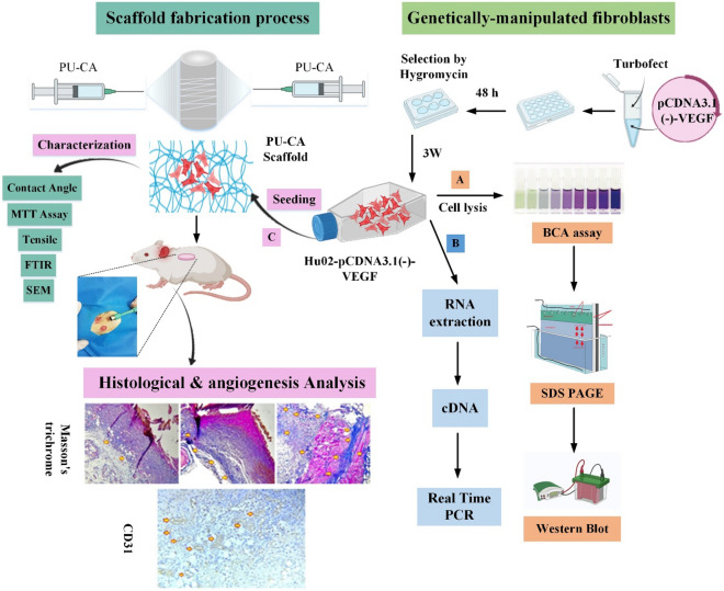
Figure 1.
Schematic illustration of in vitro and in vivo studies (Graphical Abstract). The genetically manipulated procedure was begun by constructing pcDNA3.1(-)-VEGF recombinant expression vector and then transfected into the fibroblast cells. Following the selection of fibroblast cells with hygromycin, recombinant cells were investigated in terms of VEGF expression by (A) real-time PCR and (B) western blotting methods. After the scaffold fabrication process, the mechanical, physical, and survival properties of polyurethane-cellulose acetate (PU-CA) scaffold were investigated. (C) Manipulated fibroblast cells were seeded on a PU-CA scaffold to assess the angiogenic potential in four groups containing control, PU-CA, PU-CA with fibroblast cells, and VEGF-expressing cells on different days. The healing process and angiogenesis were histopathologically evaluated by H&E, Masson’s trichrome staining, and the IHC test via the CD31 marker.

