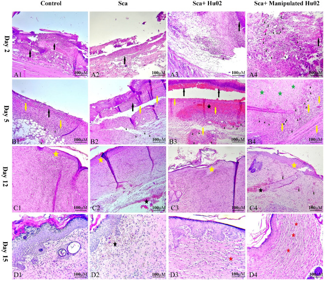
Figure 8.
H&E stained microscopic sections of wounded tissue on different days after surgery for Ctrl, Sca, Sca + Hu02, and Sca + Manipulated Hu02 groups. (A1)–(A4): Day 2 after surgery. (A1) and (A2): Tissue necrosis with high neutrophil infiltration (black arrow) and no angiogenesis. (A3): Tissue necrosis with high neutrophil infiltration (black arrow) and with mild angiogenesis as few formation of new blood vessels (arrow head). (A4): Tissue necrosis with mild neutrophil infiltration (black arrow) and remarkable angiogenesis (arrow head). (B1)–(B4): Day 5 after surgery. (B1) and (B2): Tissue necrosis with neutrophil infiltration (black arrow), mild angiogenesis (arrow head), mild fibroproliferation, and new connective tissue formation (yellow arrow). (B3): Tissue necrosis with neutrophil infiltration (black arrow), moderate angiogenesis (arrow head), hemorrhage (black star), high fibroproliferation, and new connective tissue formation (yellow arrow). (B4): Remarkable angiogenesis (arrow head), moderate fibroproliferation (yellow arrow), and new connective tissue formation and collagen deposition (green stars). (C1)–(C4): Day 12 after surgery. (C1): Reepithelialization (yellow star) and increased connective tissue. (C2): Reepithelialization (yellow star), increased connective tissue, and some area of hemorrhage (black star). (C3): Reepithelialization (yellow star) and increased connective tissue. (C4): Reepithelialization (yellow star), increased connective tissue, and some area of hemorrhage (black star). Granulation tissue and angiogenesis (arrow head) were present in some areas. (D1)–(D4): Day15 after surgery. (D1): Reduction of fibroproliferation. (D2): Reduction of fibroproliferation and the presence of subepidemal hemorrhage (black star) in some area. (D3): Reduction of fibroproliferation and beginning of scar tissue formation (red star). (D4): Reduction of fibroproliferation and remarkable scar tissue formation as large parallel bundles (red star).

