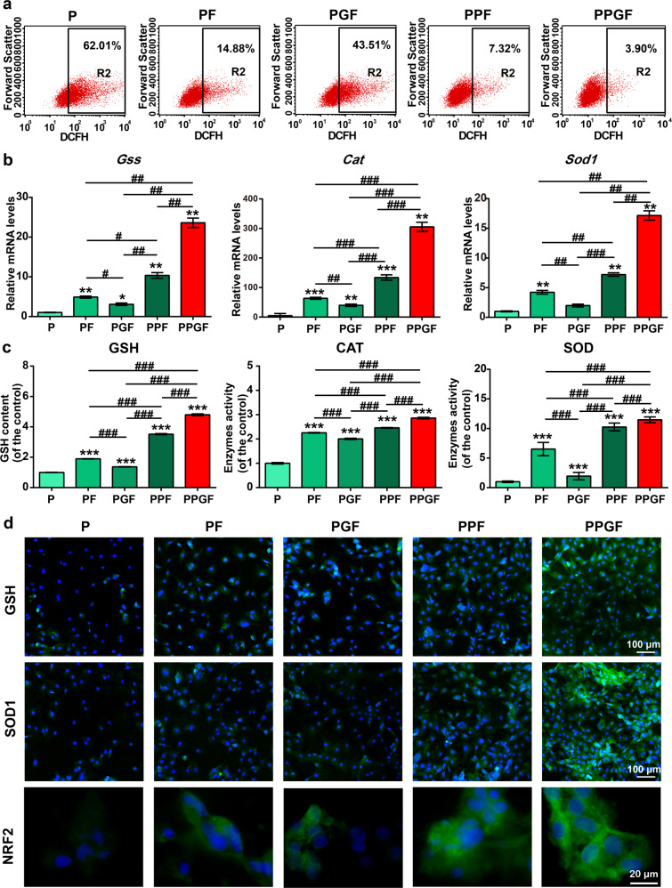
Fig. 7. Suppressive effect of nanofiber membranes on oxidative stress in Il-1β induced chondrocytes.
a ROS content in IL-1β-induced chondrocytes administrated with nanofiber membranes was evaluated by flow cytometry. b The expression of antioxidative related genes, including Cat, Gss, and Sod1, in Il-1β induced chondrocytes treated with nanofiber membranes. c Expression of antioxidative factors, including GSH, CAT, and SOD, in Il-1β-induced chondrocytes treated with nanofiber membranes, was detected by ELISA. d Expression of GSH, SOD1, and NRF2 in Il-1β-induced chondrocytes treated with nanofiber membranes was detected by immunofluorescence staining. (Scale bars, 100 µm and 20 µm). The values were presented as mean ± SD (n = 3; statistics: one-way ANOVA; *, # means p < 0.05, **, ## means p < 0.01, ***, ### means p < 0.001, * is the statistical difference compare with P and # is the statistical difference between the pairwise comparison).

