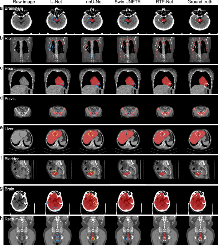
Fig. 4. Visual comparison of segmentation performance of our proposed RTP-Net, U-Net, nnU-Net, and Swin UNETR.
Segmentation is performed on eight OARs, i.e., (a) brainstem, (b) rib, (c) heart, (d) pelvis, (e) liver, (f) bladder, (g) brain, and (h) rectum. The white circles denote accurate segmentation compared to manual ground truth by four methods. The blue and yellow circles represent under-segmentation and over-segmentation, respectively.

