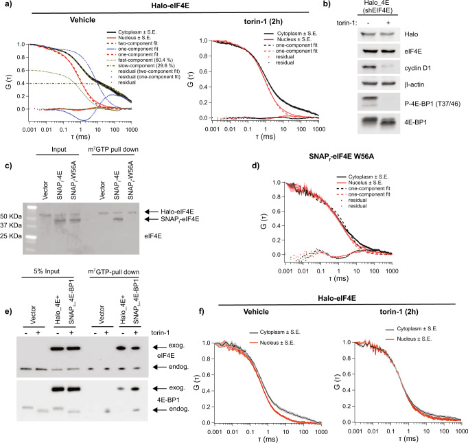
Fig. 1. Binding of exogenous Halo-eIF4E to the 5’cap is detected by FCS.
a NIH3T3 cells that express Halo-eIF4E, in which the endogenous counterpart was silenced by shRNA, were treated for 2 h with vehicle (DMSO) or 250 nM torin-1. Averaged autocorrelation curves show temporal diffusion of Halo-eIF4E molecules (ms = milliseconds) in the cytoplasm (black) and in the nucleus (red) in the indicated conditions (N = 10 ± SEM). Halo-eIF4E diffusion is slower in translating cells as compared to cells treated with torin-1. Cytoplasmic autocorrelation best fit with two components (fast – dark yellow dotted curve: τfast = 1.14 ± 0.03 ms, and slow – dash-dotted curve: τslow = 372.69 ± 10.8 ms). Percentages of slow (Dslow = 0.05 μm2/s) and fast (Dfast = 14.78 μm2/s) moving molecules are indicated. While one-component fits nuclear Halo-eIF4E well (see red curves), a one-component fit (see the blue dashed curve, with dotted fit residual) cannot adequately describe the cytoplasmic Halo-eIF4E autocorrelation curve. b Total cell lysates from the cells described in (a) were analyzed by western blotting with the indicated antibodies. 4E-BPs phosphorylation and CyclinD1 expression were significantly reduced after 2 h torin-1. c, d SNAPf-eIF4E and SNAPf-eIF4E W56A (SNAPf-W56A) were expressed in the cells described above. Total cell lysates were subjected to a cap-pull-down assay. Levels of Halo-eIF4E, SNAPf-eIF4E, and SNAPf-eIF4E W56A were detected by western blotting in input (5%) and cap-bound fractions (c) using eIF4E antibody (eIF4E). Averaged autocorrelation curves show temporal diffusion of SNAPf-eIF4E W56A in the cytoplasm (red) and in the nucleus (black) (N = 10 ± SEM). Only one fast component was detected in both cellular compartments (d). e, f NIH3T3 cells that express both Halo-eIF4E and SNAPf-4E-BP1 were treated with vehicle (DMSO) or 250 nM torin-1 for 2 h. Total cell lysates were subjected to cap-pull down assay and analyzed by western blotting in the indicated fractions Overexpression of SNAPf-4E-BP1 is sufficient to increase its binding to eIF4E on the 5’cap (e). Averaged autocorrelation curves show temporal diffusion of Halo-eIF4E in the indicated conditions (N = 10 ± SEM). SNAPf-4E-BP1 expression is sufficient to displace most of the Halo-eIF4E bound to the 5’cap (f).

