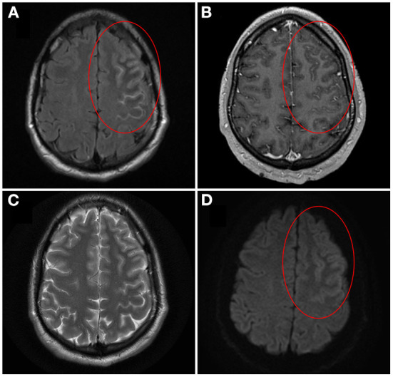
Figure 1.

Brain magnetic resonance imaging of unilateral cortical FLAIR-hyperintense Lesion in Anti-MOG-associated Encephalitis with Seizures (FLAMES)/Unilateral cerebral cortical encephalitis (UCCE). On axial T2-weighted fluid-attenuated inversion recovery (T2-FLAIR) image pre-gadolinium, cortical swelling and hyperintensity of both the left frontal cortex and adjacent sulci is seen, with hypointensity of the adjacent juxtacortical white matter [(A) circle]. On axial T1-weighted image post-gadolinium, corresponding leptomeningeal enhancement is also seen [(B) circle]. The cortical hyperintensity is not well visualized on axial T2-weighted image (C). On axial diffusion-weighted image there is brightness of the cortex with sparing of the subarachnoid space [(D) circle]. Corresponding subtle darkness on apparent diffusion coefficient map was seen, compatible with true diffusion restriction (not shown). Image adapted and re-used with permission from Springer Nature: Budhram A, Mirian A, Le C, Hosseini-Moghaddam SM, Sharma M, Nicolle MW. Unilateral cortical FLAIR-hyperintense Lesions in Anti-MOG-associated Encephalitis with Seizures (FLAMES): characterization of a distinct clinico-radiographic syndrome. J Neurol. (2019) 266(10):2481–7.
