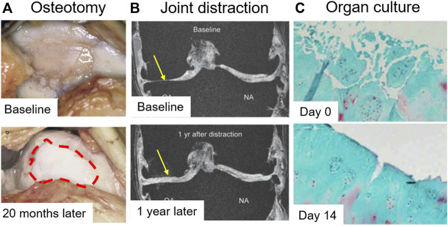
FIGURE 5.
Evidence of cartilage rebuilding in vitro and in vivo. (A) The articular surface was observed 20 months after high tibial osteotomy (HTO). The red dot circle shows neocartilage formation (Intema et al., 2011). (B) Quantitative magnetic resonance imaging (MRI) of the joint was taken after 1 year of joint distraction (Koshino et al., 2003). (C) OA cartilage was cultured for 14 days. Safranin O-fast green staining was performed (Hoshiyama et al., 2015).

