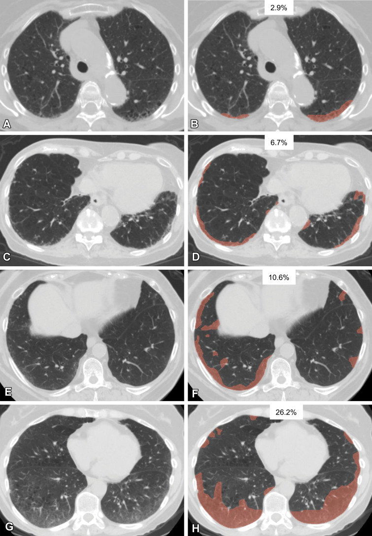
Figure 2.
Various extents of lung abnormality in four patients. Original axial CT images (A, C, E, G) and the same CT images with a segmented overlay of ILA (B, D, F, and H) are shown. (A, B) Scans in a 76-year-old woman show subtle subpleural ground-glass abnormality that accounts for 2.9% of the lung area, indicating insignificant abnormality. (C, D) Scans in an 84-year-old woman show ground-glass and reticular abnormalities that account for 6.7% of the lung area, indicating significant abnormality. (E, F) Scans in a 73-year-old woman show faint ground-glass abnormality that accounts for 10.6% of the lung area, indicating significant abnormality. (G, H) Scans in a 60-year-old woman show widespread ground-glass abnormality that affects 26.2% of the lung area, indicating significant abnormality. The segmentations shown were performed manually by using 3D Slicer software (Brigham and Women’s Hospital, Harvard Medical School, Boston, MA).

